Por favor, use este identificador para citar o enlazar este ítem:
https://hdl.handle.net/11000/38115Registro completo de metadatos
| Campo DC | Valor | Lengua/Idioma |
|---|---|---|
| dc.contributor.author | Frutos-Rincón, Laura | - |
| dc.contributor.author | Gómez Sánchez, José Antonio | - |
| dc.contributor.author | Íñigo-Portugués, Almudena | - |
| dc.contributor.author | Acosta, M. Carmen | - |
| dc.contributor.author | Gallar, Juana | - |
| dc.contributor.other | Departamentos de la UMH::Fisiología | es_ES |
| dc.date.accessioned | 2025-11-11T12:18:41Z | - |
| dc.date.available | 2025-11-11T12:18:41Z | - |
| dc.date.created | 2022-03 | - |
| dc.identifier.citation | Int J Mol Sci. 2022 Mar 10;23(6):2997 | es_ES |
| dc.identifier.issn | 1422-0067 | - |
| dc.identifier.issn | 1661-6596 | - |
| dc.identifier.uri | https://hdl.handle.net/11000/38115 | - |
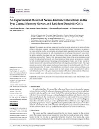
| dc.description.abstract | The cornea is an avascular connective tissue that is crucial, not only as the primary barrier of the eye but also as a proper transparent refractive structure. Corneal transparency is necessary for vision and is the result of several factors, including its highly organized structure, the physiology of its few cellular components, the lack of myelinated nerves (although it is extremely innervated), the tightly controlled hydration state, and the absence of blood and lymphatic vessels in healthy conditions, among others. The avascular, immune‐privileged tissue of the cornea is an ideal model to study the interactions between its well‐characterized and dense sensory nerves (easily accessible for both focal electrophysiological recording and morphological studies) and the low number of resident immune cell types, distinguished from those cells migrating from blood vessels. This paper presents an overview of the corneal structure and innervation, the resident dendritic cell (DC) subpopulations present in the cornea, their distribution in relation to corneal nerves, and their role in ocular inflammatory diseases. A mouse model in which sensory axons are constitutively labeled with tdTomato and DCs with green fluorescent protein (GFP) allows further analysis of the neuroimmune crosstalk under inflammatory and steady‐state conditions of the eye. | es_ES |
| dc.format | application/pdf | es_ES |
| dc.format.extent | 27 | es_ES |
| dc.language.iso | eng | es_ES |
| dc.publisher | MDPI | es_ES |
| dc.rights | info:eu-repo/semantics/openAccess | es_ES |
| dc.rights | Attribution-NonCommercial-NoDerivatives 4.0 Internacional | * |
| dc.rights.uri | http://creativecommons.org/licenses/by-nc-nd/4.0/ | * |
| dc.subject | corneal nerves | es_ES |
| dc.subject | dendritic cells | es_ES |
| dc.subject | neuro‐immune interactions | es_ES |
| dc.subject | ocular inflammation | es_ES |
| dc.subject | ocular pain | es_ES |
| dc.subject | animal models | es_ES |
| dc.title | An Experimental Model of Neuro–Immune Interactions in the Eye: Corneal Sensory Nerves and Resident Dendritic Cells | es_ES |
| dc.type | info:eu-repo/semantics/article | es_ES |
| dc.contributor.institute | Institutos de la UMH::Instituto de Neurociencias | es_ES |
| dc.relation.publisherversion | 10.3390/ijms23062997 | es_ES |
Ver/Abrir:
An Experimental Model of Neuro–Immune Interactions in the.pdf
2,14 MB
Adobe PDF
Compartir:
 La licencia se describe como: Atribución-NonComercial-NoDerivada 4.0 Internacional.