Por favor, use este identificador para citar o enlazar este ítem:
https://hdl.handle.net/11000/6997Registro completo de metadatos
| Campo DC | Valor | Lengua/Idioma |
|---|---|---|
| dc.contributor.author | Nombela Díaz, Iván | - |
| dc.contributor.author | Requena Platek, Ricardo | - |
| dc.contributor.author | Morales Lange, Byron | - |
| dc.contributor.author | CHICO GRAS, VERONICA | - |
| dc.contributor.author | Puente Marín, Sara | - |
| dc.contributor.author | Ciordia, Sergio | - |
| dc.contributor.author | Mena, María Carmen | - |
| dc.contributor.author | Coll Morales, Julio | - |
| dc.contributor.author | Pérez, Luis | - |
| dc.contributor.author | Mercado, Luis | - |
| dc.contributor.author | Ortega-Villaizan Romo, María del Mar | - |
| dc.date.accessioned | 2021-01-25T10:14:01Z | - |
| dc.date.available | 2021-01-25T10:14:01Z | - |
| dc.date.created | 2019-04-24 | - |
| dc.date.issued | 2021-01-25 | - |
| dc.identifier.issn | 2073-4409 | - |
| dc.identifier.uri | http://hdl.handle.net/11000/6997 | - |
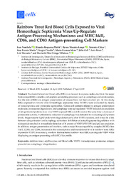
| dc.description.abstract | Nucleated teleost red blood cells (RBCs) are known to express molecules from the major histocompatibility complex and peptide-generating processes such as autophagy and proteasomes, but the role of RBCs in antigen presentation of viruses have not been studied yet. In this study, RBCs exposed ex vivo to viral hemorrhagic septicemia virus (VHSV) were evaluated by means of transcriptomic and proteomic approaches. Genes and proteins related to antigen presentation molecules, proteasome degradation, and autophagy were up-regulated. VHSV induced accumulation of ubiquitinated proteins in ex vivo VHSV-exposed RBCs and showed at the same time a decrease of proteasome activity. Furthermore, induction of autophagy was detected by evaluating LC3 protein levels. Sequestosome-1/p62 underwent degradation early after VHSV exposure, and it may be a link between ubiquitination and autophagy activation. Inhibition of autophagosome degradation with niclosamide resulted in intracellular detection of N protein of VHSV (NVHSV) and p62 accumulation. In addition, antigen presentation cell markers, such as major histocompatibility complex (MHC) class I & II, CD83, and CD86, increased at the transcriptional and translational level in rainbow trout RBCs exposed to VHSV. In summary, we show that nucleated rainbow trout RBCs can degrade VHSV while displaying an antigen-presenting cell (APC)-like profile | es |
| dc.format | application/pdf | es |
| dc.format.extent | 29 | es |
| dc.language.iso | eng | es |
| dc.rights | info:eu-repo/semantics/openAccess | es |
| dc.subject | rainbow trout | es |
| dc.subject | erythrocytes | es |
| dc.subject | red blood cells | es |
| dc.subject | VHSV | es |
| dc.subject | transcriptome | es |
| dc.subject | proteome | es |
| dc.subject | antigen presentation | es |
| dc.subject | autophagy | es |
| dc.subject | ubiquitination | es |
| dc.subject.other | 577 - Bioquímica. Biología molecular. Biofísica | es |
| dc.title | Rainbow Trout Red Blood Cells Exposed to Viral Hemorrhagic Septicemia Virus Up-Regulate Antigen-Processing Mechanisms and MHC I&II, CD86, and CD83 Antigen-presenting Cell Markers | es |
| dc.type | info:eu-repo/semantics/article | es |
| dc.contributor.institute | Instituto de Biología Molecular y Celular | es |
Ver/Abrir:
8-Nombela et al_2019_Rainbow Trout Red Blood Cells Exposed to VHSV Up-Regulate Antigen-presenting Cell Markers_cells.pdf
6,47 MB
Adobe PDF
Compartir:
 La licencia se describe como: Atribución-NonComercial-NoDerivada 4.0 Internacional.