Por favor, use este identificador para citar o enlazar este ítem:
https://hdl.handle.net/11000/31354Registro completo de metadatos
| Campo DC | Valor | Lengua/Idioma |
|---|---|---|
| dc.contributor.author | Belda, José I. | - |
| dc.contributor.author | Artola, Alberto | - |
| dc.contributor.author | García Manzanares, María Dolores | - |
| dc.contributor.author | Ferrer, Consuelo | - |
| dc.contributor.author | Haroun, Hazem E. | - |
| dc.contributor.author | hassanein, ahmed | - |
| dc.contributor.author | Baeyens, Vincent | - |
| dc.contributor.author | Munoz, Gonzalo | - |
| dc.contributor.author | Alió, Jorge L. | - |
| dc.contributor.other | Departamentos de la UMH::Producción Vegetal y Microbiología | es_ES |
| dc.date.accessioned | 2024-02-09T10:41:29Z | - |
| dc.date.available | 2024-02-09T10:41:29Z | - |
| dc.date.created | 2004-11 | - |
| dc.identifier.citation | Journal of Cataract and Refractive Surgery, vol. 31 (2005) | es_ES |
| dc.identifier.issn | 0886-3350 | - |
| dc.identifier.issn | 1873-4502 | - |
| dc.identifier.uri | https://hdl.handle.net/11000/31354 | - |
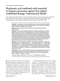
| dc.description.abstract | Purpose: To evaluate the protective properties of combined sodium hyaluronate2% and mannitol 0.5% (Visiol) on the corneal endothelium in the presence of oxidative stress induced by hydrogen peroxide (H2O2). Setting: Instituto Oftalmolo´ gico de Alicante, Universidad Miguel Herna´ ndez, Alicante, Spain. Methods: This was an exploratory randomized controlled parallel-group, maskedassessor study of 3 sodium hyaluronate-based ophthalmic viscosurgical devices (OVDs): Visiol, Healon (sodium hyaluronate 1%), and Viscoat (sodium hyaluronate 3%–chondroitin sodium 4%). The OVDs were tested for protective effects on the endothelium following oxidative stress induced by H2O2 at increased concentrations: control (lactated Ringer’s solution), 1mM, 10mM, and 100 mM. Groups without OVD were used as controls at the same concentrations of peroxide. Each animal received the same treatment in both eyes (10 eyes per group). Endothelial cell lesion was assessed using the Janus green photometry absorbance technique. Results: At 10 mM peroxide concentration, the value of endothelial cell lesion was significantly lower in the Visiol (16.8%, P Z .0056), Healon (22.2%, P Z .0302), and Viscoat (21.6%, PZ .0336) groups than in the control group (29.4%, no OVD). There was a trend in favor of Visiol to more efficiently reduce cell lesions of the endothelium, than Healon (PZ .055) and Viscoat (P Z .1013). Values of endothelial cell lesion at peroxide concentrations of 1 mM and 100 mM showed the same trends than those observed at 10 mM. Conclusions: All of the OVDs tested efficiently reduced endothelial lesions against free radicals compared with the control group in which no OVD was used. The following sequence for the efficacy of endothelial cell protection was established: Visiol O Viscoat O Healon O no OVD. | es_ES |
| dc.format | application/pdf | es_ES |
| dc.format.extent | 6 | es_ES |
| dc.language.iso | eng | es_ES |
| dc.publisher | Williams & Wilkins | es_ES |
| dc.rights | info:eu-repo/semantics/openAccess | es_ES |
| dc.rights | Attribution-NonCommercial-NoDerivatives 4.0 Internacional | * |
| dc.rights.uri | http://creativecommons.org/licenses/by-nc-nd/4.0/ | * |
| dc.title | Hyaluronic acid combined with mannitol to improve protection against free-radical endothelial damage: Experimental Model | es_ES |
| dc.type | info:eu-repo/semantics/article | es_ES |
| dc.relation.publisherversion | https:// doi.org/10.1016/j.jcrs.2004.11.055 | es_ES |
Ver/Abrir:
2004 Hyaluronic acid combined with mannitol to improve protection against free-radical endothelial damage experimental model.pdf
159,04 kB
Adobe PDF
Compartir:
 La licencia se describe como: Atribución-NonComercial-NoDerivada 4.0 Internacional.