Título :
FXR modulates the gut-vascular barrier by regulating the entry sites for bacterial translocation in experimental cirrhosis |
Autor :
Sorribas, Marcel
Jakob, Manuel O.
Yilmaz, Bahtiyar
Li, Hai
Stuzt, David
Noser, Yannik
de Gottardi, Andrea
Moghadamrad, Sheida 
Hassan, Moshin
Albillos, Agustin
Francés, Ruben
Juanola, Oriol
Spadoni, Ilaria
Rescigno, Maria
Wiest, Reiner |
Editor :
Elsevier |
Departamento:
Departamentos de la UMH::Medicina Clínica |
Fecha de publicación:
2019-12 |
URI :
https://hdl.handle.net/11000/31335 |
Resumen :
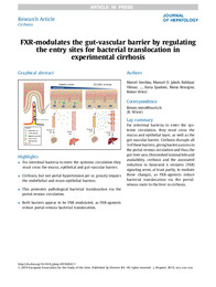
Background & aims: Pathological bacterial translocation (PBT) in cirrhosis is the hallmark of spontaneous bacterial infections, increasing mortality several-fold. Increased intestinal permeability is known to contribute to PBT in cirrhosis, although the role of the mucus layer has not been addressed in detail. A clear route of translocation for luminal intestinal bacteria is yet to be defined, but we hypothesize that the recently described gut-vascular barrier (GVB) is impaired in experimental portal hypertension, leading to increased accessibility of the vascular compartment for translocating bacteria.
Materials: Cirrhosis was induced in mouse models using bile-duct ligation (BDL) and CCl4. Pre-hepatic portal-hypertension was induced by partial portal vein ligation (PPVL). Intestinal permeability was compared in these mice after GFP-Escherichia coli or different sized FITC-dextrans were injected into the intestine.
Results: Healthy and pre-hepatic portal-hypertensive (PPVL) mice lack translocation of FITC-dextran and GFP-E. coli from the small intestine to the liver, whereas BDL and CCl4-induced cirrhotic mice demonstrate pathological translocation, which is not altered by prior thoracic-duct ligation. The mucus layer is reduced in thickness, with loss of goblet cells and Muc2-staining and expression in cirrhotic but not PPVL mice. These changes are associated with bacterial overgrowth in the inner mucus layer and pathological translocation of GFP-E. coli through the ileal epithelium. GVB is profoundly altered in BDL and CCl4-mice with Ileal extravasation of large-sized 150 kDa-FITC-dextran, but only slightly altered in PPVL mice. This pathological endothelial permeability and accessibility in cirrhotic mice is associated with augmented expression of PV1 in intestinal vessels. OCA but not fexaramine stabilizes the GVB, whereas both FXR-agonists ameliorate gut to liver translocation of GFP-E. coli.
Conclusions: Cirrhosis, but not portal hypertension per se, grossly impairs the endothelial and muco-epithelial barriers, promoting PBT to the portal-venous circulation. Both barriers appear to be FXR-modulated, with FXR-agonists reducing PBT via the portal-venous route.
Lay summary: For intestinal bacteria to enter the systemic circulation, they must cross the mucus and epithelial layer, as well as the gut-vascular barrier. Cirrhosis disrupts all 3 of these barriers, giving bacteria access to the portal-venous circulation and thus, the gut-liver axis. Diminished luminal bile acid availability, cirrhosis and the associated reduction in farnesoid x receptor (FXR) signaling seem, at least partly, to mediate these changes, as FXR-agonists reduce bacterial translocation via the portal-venous route to the liver in cirrhosis.
|
Palabras clave/Materias:
FXR
Gut-liver axis
Gut-vascular barrier
Intestinal permeability
Liver cirrhosis
Mucus
Portal hypertension |
Tipo de documento :
info:eu-repo/semantics/article |
Derechos de acceso:
info:eu-repo/semantics/openAccess
Attribution-NonCommercial-NoDerivatives 4.0 Internacional |
DOI :
https://doi.org/10.1016/j.jhep.2019.06.017 |
Publicado en:
Journal of Hepatology. 2019 Dec;71(6):1126-1140 |
Aparece en las colecciones:
Artículos Medicina Clínica
|
 La licencia se describe como: Atribución-NonComercial-NoDerivada 4.0 Internacional.