Please use this identifier to cite or link to this item:
https://hdl.handle.net/11000/28980Full metadata record
| DC Field | Value | Language |
|---|---|---|
| dc.contributor.advisor | Canals Gamoneda, Santiago | - |
| dc.contributor.author | Pérez-Cervera, Laura | - |
| dc.contributor.other | Instituto de Neurociencias | es_ES |
| dc.date.accessioned | 2023-03-17T09:59:29Z | - |
| dc.date.available | 2023-03-17T09:59:29Z | - |
| dc.date.created | 2022-07-04 | - |
| dc.identifier.other | 1718 | - |
| dc.identifier.uri | https://hdl.handle.net/11000/28980 | - |
| dc.description.abstract | Alcohol addiction is a health problem that causes millions of deaths worldwide every year. However, the understanding of how alcohol-induced brain alterations lead to addiction remains limited and thus, effective targets for treatment are elusive. The diversity of damage it can cause to the brain has been studied in depth, but is very heterogeneous between individuals. Vulnerability to relapse, which is maximal in the early stages of the disease, remains an unsolved enigma. Typical characteristics of alcoholic patients, such as lack of cognitive control or behavioral inflexibility, could trigger relapse, but little is known about the possible mechanisms underlying them. A significant problem in clinical studies on AUD is the high variability of the results obtained. This variability is partly due to the variety of personal trajectories in alcohol use and abuse and genetic factors, but also to the high number of possible comorbidities associated with alcoholism. These include poly-substance use (tobacco, cannabis, cocaine, etc.) and pharmacological treatments to alleviate the multiple associated symptoms. In this thesis work, I have taken advantage of well-controlled animal models to study the transformations that occur from a control state, naive to alcohol consumption, to excessive consumption and subsequent abstinence. At the same time, several cohorts of humans have been studied, using at all times the same magnetic resonance imaging (MRI) modalities for the study of brain alterations. This design allows to establish causal relationships with alcohol consumption in the animal models, avoiding comorbidities and other confounding factors, without moving away from translation to the clinic (thanks to the convergent set of brain imaging tools used). The first objective was to study the evolution of brain structure during the early phases of abstinence. We used a rat model with a genetic predisposition to consumption, and two cohorts of AUD patients at different times of early abstinence. Brain alterations were studied using diffusion tensor magnetic resonance imaging (DTI), a non-invasive technique with diagnostic value, which informs about water diffusion in tissues. From this information we can infer microstructural alterations. We found that both rats and humans showed comparable microstructural alterations in the white matter (WM) in the early stages of withdrawal. Unexpectedly, we were able to demonstrate that these alterations progressed throughout the period of abstinence studied. Taken together, these findings suggested the existence of an underlying biological process that triggers the progression of WM alterations in the absence of alcohol. In a follow-up study, using the same cohorts of patients, we made a detailed study of the effect size of the alterations found, distinguishing between brain tracts. This study revealed that the most vulnerable tract in alcoholic patients was the fimbria/fornix, which communicates the hippocampus with the prefrontal cortex. Considering that this communication is fundamental for both learning and memory functions, as well as for emotional regulation (together with the amygdala) and cognitive flexibility, we argue that fimbria/fornix dysfunction in the early phase of abstinence could contribute to some key symptoms commonly observed in patients. Consequently, we decided to study in more detail and using again an animal model, the structure and functionality of the fimbria/fornix after alcohol withdrawal. On this occasion, we used a rat model that has demonstrated alcohol dependence, based on wild type animals, known as the postdependent or chronic intermittent exposure model. In addition to diffusion MRI, we used immunohistological and electrophysiological techniques. The results indicated a microstructural alteration in the tract, measured both as a decrease in myelin fraction quantified with MRI, and a decrease in myelin basic protein in postmortem histological examinations. In addition, simultaneous electrophysiological recordings demonstrated reduced efficiency in the directed connectivity from the hippocampus to the prefrontal cortex, together with an increased excitation/inhibition balance in the hippocampus. Taken together, these results suggest that an alcohol-driven mechanism could be damaging the brain microstructure even in the absence of alcohol. Although the underlying mechanism has not been studied, in the case of fimbria/fornix it corresponds to a demyelinating process. The functional consequence of the latter is the partial disconnection of the hippocampus and the prefrontal cortex. We propose that this alteration could be triggering some of the symptoms that characterize alcoholic patients, such as memory deficits and reduced cognitive flexibility, which condemns patients to compulsively repeat behaviors associated with alcohol consumption. | es_ES |
| dc.format | application/pdf | es_ES |
| dc.format.extent | 114 | es_ES |
| dc.language.iso | eng | es_ES |
| dc.publisher | Universidad Miguel Hernández de Elche | es_ES |
| dc.rights | info:eu-repo/semantics/openAccess | es_ES |
| dc.rights.uri | http://creativecommons.org/licenses/by-nc-nd/4.0/ | * |
| dc.subject | Neurofisiología | es_ES |
| dc.subject.other | CDU::6 - Ciencias aplicadas::61 - Medicina::616 - Patología. Medicina clínica. Oncología::616.8 - Neurología. Neuropatología. Sistema nervioso | es_ES |
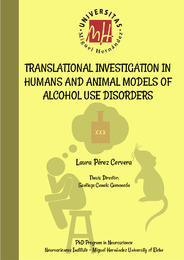
| dc.title | Translational investigation in humans and animal models of alcohol use disorders | es_ES |
| dc.type | info:eu-repo/semantics/doctoralThesis | es_ES |

View/Open:
2.Tesis_FULL_SC (1) Pérez Cervera, Laura.pdf
20,62 MB
Adobe PDF
Share:
.png)
